Figure 6.

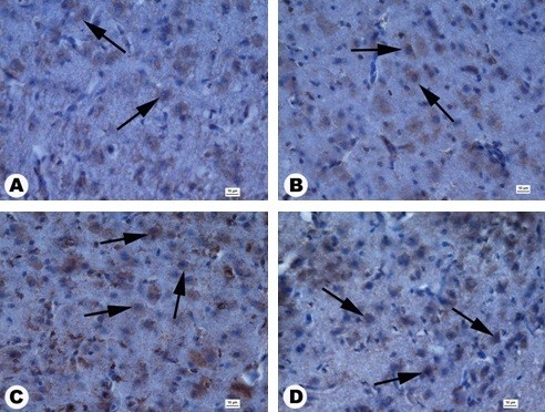
Cross sections of a granular layer in the cerebral cortex by anti-caspase-3 staining. (A) Control, (B) 1 μg/ml, (C) 10 μg/ml, (D) 20 μg/ml. Anti-caspase-3-positive cells (arrows). Scale bars 10 μm.

Cross sections of a granular layer in the cerebral cortex by anti-caspase-3 staining. (A) Control, (B) 1 μg/ml, (C) 10 μg/ml, (D) 20 μg/ml. Anti-caspase-3-positive cells (arrows). Scale bars 10 μm.