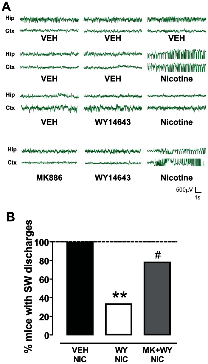
Figure 2. The PPARα agonist WY14643 suppresses nicotine-induced spike-wave activity.
(A) Representative traces of EEG recordings from hippocampal (Hip) and sensorimotor cortical (Ctx) electrodes chronically implanted in mice. Following the administration of vehicle (VEH) and 10 mg/kg nicotine, bursts of synchronous spike-wave (SW) activity with high-amplitude and low-frequency (most in the delta rhythm range) were recorded. This activity was suppressed when animals were pretreated with the PPARα agonist WY14643 (WY, 80 mg/kg), which per se did not change baseline EEG activity. The PPARα antagonist MK886 (MK, 3 mg/kg, i.p.) restored nicotine-induced SW discharges. (B) The graph shows the percentage of mice presenting SW discharges following the three treatment protocols. Vehicles treated mice did show SW activity, whereas 100% of nicotine treated mice displayed bursts of SW activity. The effects of nicotine were blocked in the majority of WY treated mice, since SW burst were recorded only in 33% of treated animals (**P<0.01 vs. vehicle, Fisher’s test). Conversely, when MK was administered 15 before WY nicotine-induced SW activity was recorded in 78% of MK+WY treated mice (#P<0.05 vs. WY, Fisher’s test).

