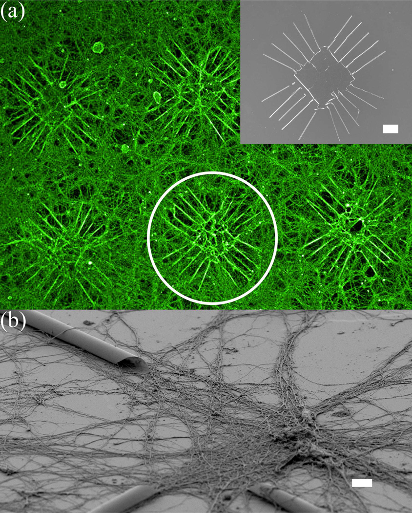
Figure 3.
Fluorescent-microscopy and SEM images of cortical neurons cultured on a Si wafer with Si/SiGe tubes. (a) Pseudo-colored fluorescent image of neurons growing extensively on an array of patterns, which consist of multiple extending tubes on each side. Inset is a SEM picture of the pattern. The fluorescent image is taken using antibody to tubulin that heavily labels dendrites and axons. Semiconductor structures are invisible in the fluorescent image, but are outlined by the contrast of surrounding fluorescently-labeled neurons and processes, as shown inside the white circle; (b) SEM image of neurons at an intersection of several tubes. Neurons appear to grow processes in the entire area, although more concentrated in the vicinity of the tubes. Scale bar is 100 µm for (a) and 10 µm for (b).

