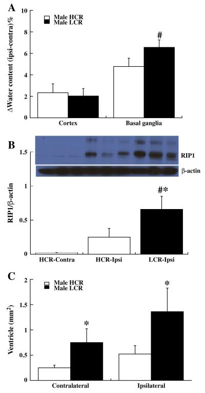
Fig. 1.
Graphs showing: (A) Brain edema (% water content, ipsiminus contralateral) in cortex and basal ganglia of HCR and LCR male rats at day 3 after ICH. Values are means±SD, n=6, #p<0.01 vs. HCR by student T test. (B) RIP1 protein levels in the ipsiand contralateral basal ganglia of male HCRs and the ipsilateral basal ganglia of male LCRs at day 7 after ICH. Values are means±SD, n=4, #p<0.01 and *p<0.05 vs. HCRs. (C) Lateral ventricular areas in HCR and LCR male rats at day 28 after ICH. Values are means±SD, *p<0.05 vs. HCR.

