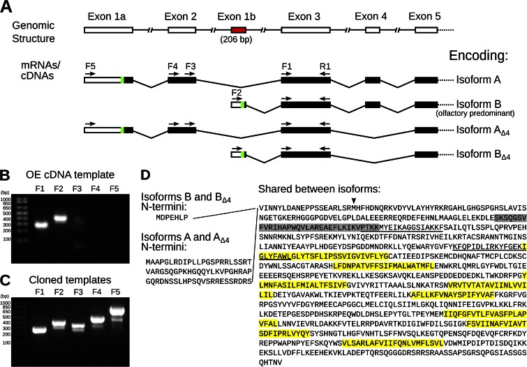
Figure 1.
Characterization of Ano2 mRNA variants present in mouse olfactory epithelium. (A) Diagram summarizing the 5′ Ano2 exon splicing structure. The green sections indicate the most 5′ AUG translation start codons, and the subsequent black bars indicate predicted protein-coding sequence. The variants containing exons 1a and 2 are less abundant in the olfactory epithelium than the variants containing the newly discovered exon 1b (red), as determined in B and C. The five forward (F1–F5) and one reverse (R1) PCR primer–binding sites are indicated as arrows. Given two alternative 5′ ends and alternative splicing of exon 4, these mRNA variants may encode up to four ANO2 isoforms, named on the right. (B and C) Ethidium bromide–stained agarose gels showing RT-PCR products with primers specific for each 5′ variant of Ano2. Primer F1 is universal for both variants, F2 is specific for exon 1b, F3 and F4 are specific for exon 2, and F5 is specific for exon 1a. The reverse primer (R1) was the same for all PCR reactions. The expected PCR product sizes are 267, 409, 350, 511, and 830 bp, respectively. Quantification of the bands by densitometry is shown in Fig. S1 C. (B) PCR template was olfactory epithelium cDNA. (C) Positive control PCR using cloned templates of equal amounts. (D) Corresponding amino acid sequence of the olfactory ANO2 isoforms. The amino acids encoded by exon 4 are highlighted in gray. Predicted transmembrane domains are highlighted in yellow. Putative CaM-binding sites are underlined (Yap et al., 2000; Tian et al., 2011). The start site used in Stephan et al. (2009) is indicated by an arrowhead.

