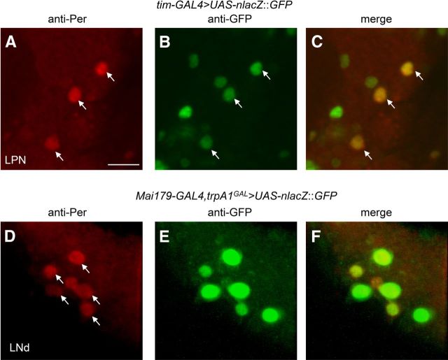
Figure 6.
Testing for coexpression of anti-Per with reporters in LPNs and LNds. A–C, The tim-GAL4 reporter was expressed in LPNs. A brain was dissected from UAS-nlacZ::GFP/tim-GAL4 flies at ZT21, after the flies were exposed to 18 h thermophase (29°C)/6 h cryophase (18°C) cycles in the dark for 6 d. The brain was stained with anti-Per and anti-GFP. A, anti-Per (red). The arrows point to LPN neurons that were Per positive. B, anti-GFP (green). C, The merged image from A and B. D–F, Colabeling with anti-Per (red) and anti-GFP (green) from UAS-mCD8::GFP/Mai179-GAL4;trpA1GAL4/+ flies at ZT21 after entraining flies under TC cycles for 6 d in the dark. D, anti-Per (red). A total of six LNds are labeled. The arrows point to LNds that were Per and GFP positive in merged image in F. E, Anti-GFP (green). F, The merged image from D and E. Scale bar, 10 μm.

