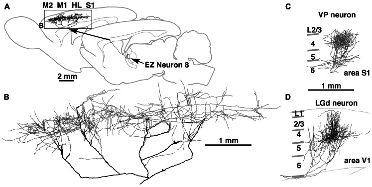
FIGURE 3.
Axonal arborization of an EZ neuron in comparison with that of primary sensory thalamic neurons in rats. The axons of EZ neurons were distributed very widely, often in the cortical area spanning more than 5 mm (A,B). This distribution is in sharp contrast to those of sensory thalamic neurons, such as neurons in the VP and dorsal lateral geniculate nucleus (LGd). The axonal arborization of VP and LGd neurons was concentrated to the middle layers of a cortical region with the size of a single column (C,D). Modified with permission from Figure 8 of Kuramoto et al. (2009) (A,B) and Figure 3 of Furuta et al. (2011) (C). The axonal arborization of a single LGd neuron in area V1 (D) was labeled and illustrated with the same method as that in Kuramoto et al. (2009) by Nakamura and Kaneko.

