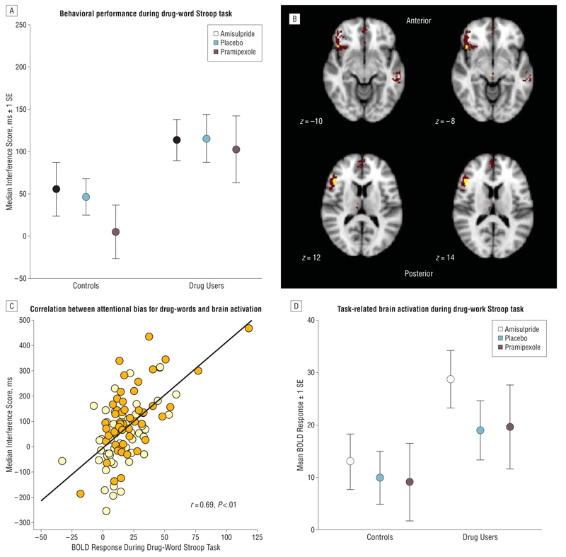
Figure 2.
Behavioral performance during the drug-word Stroop paradigm and task-related activation of a frontocerebellar system associated with attentional bias for drug words (in all participants). A, Drug users showed a significant attentional bias for drug-related words, as reflected in higher attentional interference scores compared with the healthy comparison volunteers. Attentional bias was measured by each volunteer’s median response latency of correctly identified colors of drug-related words minus the median response latency of correctly identified colors of matched neutral words. Pramipexole was given as pramipexole dihydrochloride. B, The red voxels indicate brain regions activated by the contrast between drug-related words and neutral words; yellow voxels indicate brain regions within this system where activation was positively correlated with attentional interference scores on the drug-word Stroop task. C, Scatterplot of median attentional interference score (y-axis) vs functional activation of the brain regions associated with attentional bias for drug words (x-axis), which are the left ventral prefrontal cortex (Montreal Neurological Institute coordinates [x, y, z] in millimeters: −46, 26, 12; −44, 22, −8; and −40, 6, 30) and right cerebellum (22, −80, −40). The spatial coordinates refer to the peak voxel where the effect size is greatest. BOLD indicates blood oxygen level dependent. D, Comparison of mean task-related (BOLD) activation in brain regions associated with attentional bias between stimulant-dependent individuals and healthy volunteers. Stimulant users show overactivation in the left ventral prefrontal cortex and right cerebellum compared with controls.

