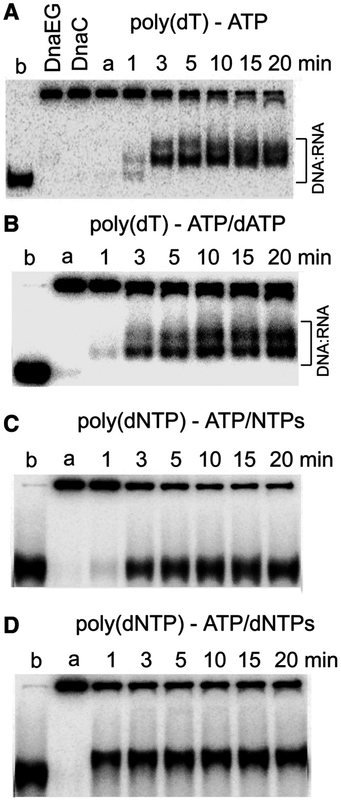
Figure 8.

Primer formation during helicase assays. (A) Coupled helicase–primase–polymerase assays were carried out with the poly(dT)-tailed DNA substrate as described in Figure 6B, but in the presence of DnaEBs (83.3 nM). The shift of the displaced oligonucleotide appears earlier than in equivalent reactions in the absence of DnaEBs (compare with Figure 6B), indicating the DnaEBs stimulates primer synthesis. (B) Similar reactions as in panel A were carried out, but this time in the presence of dATP (0.5 mM). Similar shifts of the displaced oligonucleotide were observed compared with panel A. (C) Coupled helicase–primase assays were carried out with the random sequence tail DNA substrate, which has a single 5′-d(TTTT) site on the 5′ tail, in the presence of rNTPs (0.5 mM each). No shift of the displaced oligonucleotide was observed. (D) Coupled helicase–primase–polymerase assays were carried out as described in panel C, but this time in the presence of dNTPs (0.5 mM each; no rNTPs). A clear well-defined shift of the displaced oligonucleotide was apparent from the first time point (1 min), suggesting the formation of very small RNA primers that were extended further by DnaEBs.
