Figure 5.
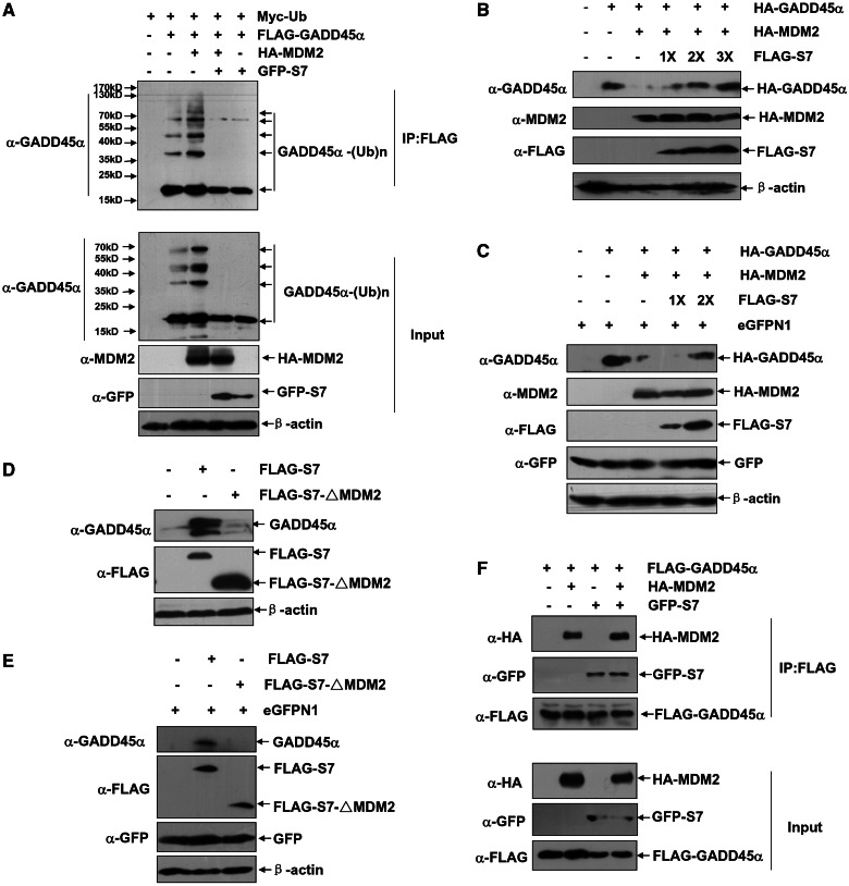
S7 attenuates MDM2-mediated GADD45α ubiquitination and degradation. (A) HepG2 cells were transfected with Myc–Ub expression plasmid in combination of FLAG–GADD45α, HA–MDM2 or GFP–S7 constructs as indicated. Then the ubiquitination of GADD45α was detected with anti-GADD45α antibody. (B) HepG2 cells were left untreated or transfected with HA–GADD45α construct (0.25 µg) with or without combination of HA–MDM2 expression plasmid (1.25 µg) and increasing amount of FLAG–S7 construct (1.25, 2.5 and 3.75 µg, respectively). Then the levels of GADD45α were detected. (C) H1299 cells were left untreated or transfected with HA–GADD45α construct (0.5 µg) with or without combination of HA–MDM2 expression plasmid (1.5 µg) and increasing amount of FLAG–S7 construct (1.5 and 3.0 µg, respectively). Then the levels of GADD45α were detected. (D and E) HepG2 (D) or H1299 (E) cells were transfected with equal amount of FLAG–S7 or FLAG–S7–ΔMDM2 constructs (3 µg), and then the levels of the endogenous GADD45α were detected. (F) 293 T cells were transfected with combination of FLAG–GADD45α, HA–MDM2 or GFP–S7 constructs as indicated. The cells were subjected to MG132 (10 µM) treatment for 4 h before harvesting. Then cell lysate was immunoprecipitated with anti-FLAG antibody, and the immunoprecipitants were probed with anti-FLAG, anti-GFP or anti-MDM2 antibodies.

