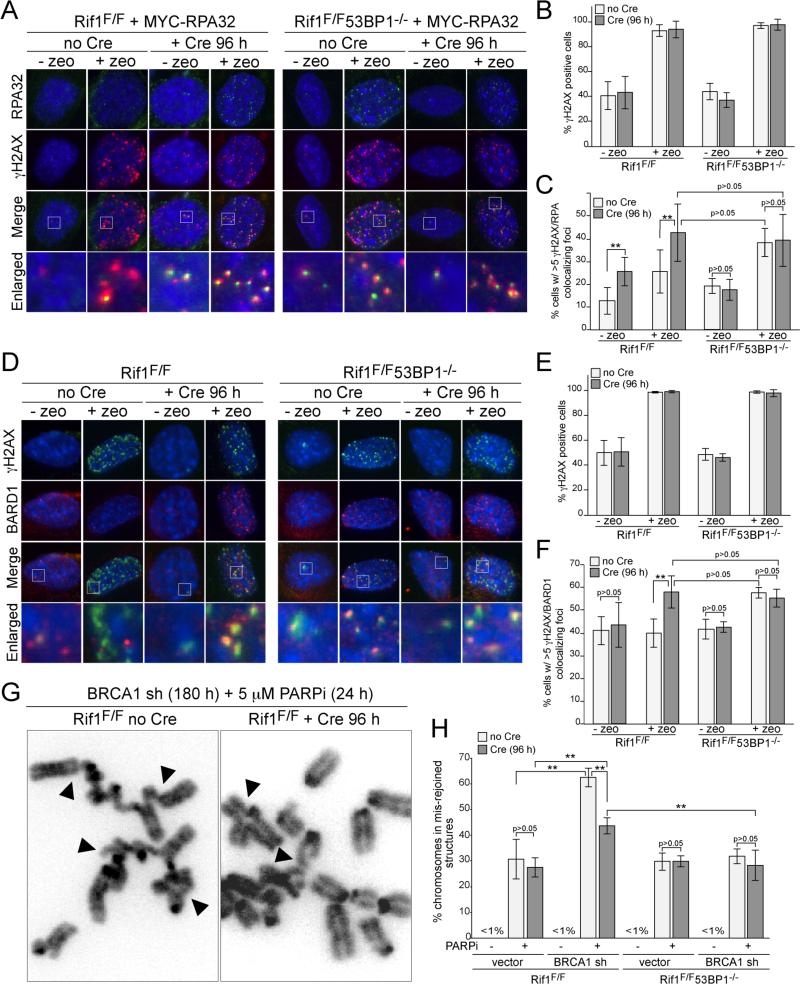
Figure 4. Rif1 inhibits resection at DSBs and promotes radial formation.
(A) IF for γ-H2AX (red) and MYC-RPA32 (green) in Cre-treated SV40-LT immortalized Rif1F/F and Rif1F/F53BP1-/- cells expressing MYC-RPA32 treated with zeocin (100 ug/ml, for 1 h; 2 h prior to analysis). (B) Percentage of γ-H2AX positive cells in experiments as in (A). (C) Percentage of cells (as in A) scored positive when containing at least five γ-H2AX foci co-localizing with RPA. (D) IF for γH2AX (green) and BARD1 (red) in Rif1F/F and Rif1F/F53BP1-/- MEFs. Cells and treatment as in (A). (E) Percentage of γ-H2AX positive cells in experiments in (D). (F) Percentage of cells in (D) containing >5 BARD1/γ-H2AX colocalizing foci. (G) Examples of mis-rejoined and radial chromosomes (arrowheads) in BRCA1sh/PARPi-treated Rif1F/F cells with or without Cre. (H) Percentages of chromosomes that are mis-rejoined in the indicated genotypes and treatments. Data in (B,C), (E,F) and (H) are means of 3-5 experiments ±SDs. ** indicates p values <0.05 (two-tailed paired Student's t-test).

