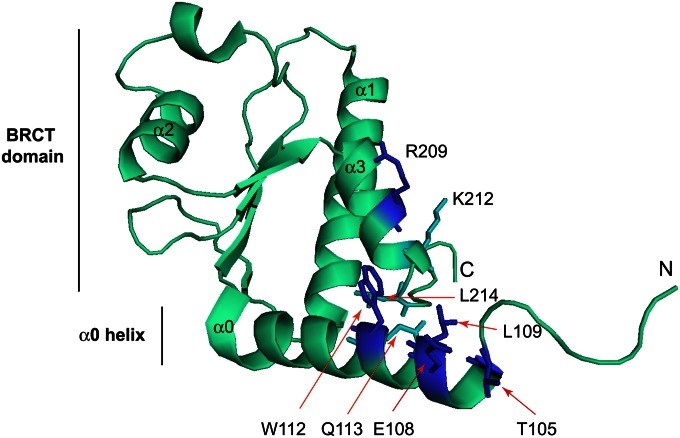
Figure 8.
Structural representation of the budding yeast BRCT domain and a preceding α-helix (α0) (both in cyan) using PDB coordinates 3QBZ (Matthews et al. 2012). The helices within the BRCT domain are numbered α1, α2, and α3. Five residues that are important for interaction with Rad53 (T105, E108, L109, W112, R209) are colored blue. L214 and Q113, which form a hydrophobic surface with W112, are colored in cyan. K212 is also colored in cyan but is not important for the Rad53 interaction.

