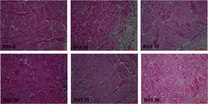
Figure 3.
Recellularization and revascularization of implanted PADM materials over time. H&E staining showed cellular content from days 5 to 30. Scale bar is 100 µm. Histology scores are summarized in Table 3.
PADM: porcine-derived acellular dermal matrix; H&E: hematoxylin and eosin.

