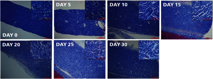
Figure 4.

Low- and high-magnification images of PADM firm materials stained with trichrome blue. Trichrome staining on day 0 showed dense collagen bundles that progressively loosened through day 25. Increased separation and white space between and across collagen bundles were noted from days 0 to 20. Collagen staining between days 20 and 30 showed a substantial increase in the density of bundles. Decreased white space and bundle separation were observed from days 20 to 30. Subtle differences in the intensity of blue staining, meant to differentiate newer collagen (lighter blue) from older collagen (darker blue), was also noted from days 10 to 30 between existing collagen bundles. These slides were not scored.
PADM: porcine-derived acellular dermal matrix.
