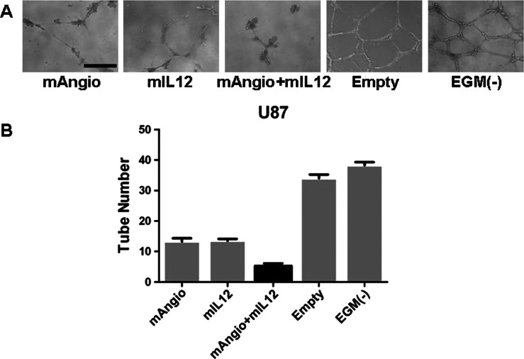
Figure 1.
Antiangiogenic effect of supernatants collected from virus-infected U87 cells in vitro. (A) Representative images of tubular structures formed by HUVECs when grown on a matrigel substrate with supernatants from infected U87 cells. The bar indicates 0.5 mm. (B) The number of tubes counted when HUVECs were grown in the presence of supernatants from U87 cells infected with G47Δ-Empty (Empty; 35.25 ± 0.85), G47Δ-mAngio (mAngio; 13.25 ± 1.11), G47Δ-mIL12 (mIL12; 13.50 ± 0.65), G47Δ-mAngio + G47Δ-mIL12 (mAngio + mIL12; 5.25 ± 0.85), or EGM (-; 38.25 ± 1.12). The bars indicate average number of tubes ± SD. Statistically significant differences were observed for Empty versus mAngio, P < .0001; Empty versus mIL12, P < .0001; mAngio versus mAngio + mIL12, P = .0012, mIL12 versus mAngio + mIL12, P = .0006; EGM(-) versus mAngio + mIL12, P < .0001; EGM(-) versus mAngio, P < .0001; EGM(-) versus mIL12, P < .0001.

