Figure 1.

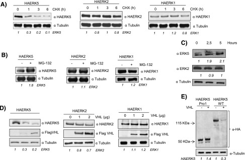
HA-ERK5 is degraded through the proteasome. (A) Cos7 cells were transfected with 0.5 µg of HA-ERK5, HA-ERK2, and HA-ERK1 and, 36 hours later, treated with 100 µM cycloheximide for the indicated times. Then, 30 µg of total cell lysates (TCLs) were blotted against indicated antibodies. (B) Cos7 cells were transfected as in A and treated with 20 µM MG132 for 5 hours. Then, 30 µg of TCLs were blotted against HA and tubulin. (C) Cos7 cells were treated with 20 µM MG132 for indicated times. Then, 60 µg of TCLs were blotted against ERK5, ERK2, and tubulin. (D) Cos7 cells were transfected with 0.5 µg of HA-ERK5, HA-ERK2, and HA-ERK1 plus increasing amounts of Flag-VHL. Thirty-six hours later, 30 µg of TCLs were blotted against the indicated antibodies. (E) Western blot of Cos7 cells transfected with 0.5 µg of HA-ERK5 Pro1 or HA-ERK5 WT in the presence or absence of 2 µg of Flag-VHL. Lysates were blotted against HA and tubulin as loading control. Fold variation of these experiments for each MAPK is shown at the bottom of each panel.
