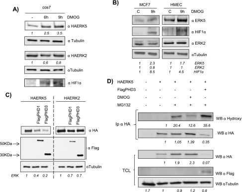
Figure 3.

ERK5 levels are regulated through a prolyl hydroxylation mechanism. (A) Cos7 cells were transfected as in Figure 1A. Thirty-six hours later, cells were treated with 1.5 mM DMOG at indicated times. TCLs were blotted against HA, HIF-1α, and tubulin. (B) Subconfluent cultures of MCF7 and HMEC cell lines were treated with 1.5 mM DMOG for 9 hours and endogenous levels of ERK5 (60 µg), ERK2 (30 µg), HIF-1α (60 µg), and tubulin (10 µg) were detected by immunoblot analysis using TCL. (C) Cos7 cells were transfected with 0.5 µg of HA-ERK5 or HA-ERK2 in the presence/absence of 2 µg of FlagPHD-1 or FlagPHD-3 and processed as in Figure 1C. (D) Cos7 cells were transfected with 0.5 µg of HA-ERK5 alone or with 2 µg of FlagPHD-3. Thirty-six hours later, cells, except control, were treated with 20 µM MG132 in the presence/absence of 1.5 mM DMOG for 12 hours. Then, extracts were collected and immunoprecipitated against HA and blotted with the indicated antibody and reblotted against HA. Thirty micrograms of TCL were blotted against HA, Flag, and tubulin. Fold variations for HA-tagged proteins or endogenous proteins in each experiment are indicated at the bottom of the panels.
