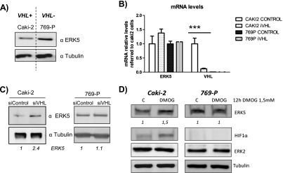
Figure 4.

VHL mediates ERK5 expression level in renal carcinoma-derived cell lines. (A) Caki-2 and 769-P cell lines were tested for ERK5 (60 µg) and tubulin (10 µg) expression by Western blot by using lysates from subconfluent cultures. (B) Levels of RNA ERK5 were analyzed by qRT-PCR in Caki-2 and 769-P cells 48 hours after transfection of control or VHL siRNA cells. (C) Caki-2 and 769-P were transfected as in B, and 60 hours later, ERK5 protein levels were analyzed by using 60 µg of cell lysates. Tubulin (10 µg) was used as loading control. (D) Subconfluent cultures of Caki-2 and 769-P cell lines were treated with 1.5 mM DMOG for 9 hours. Then, TCLs were collected and 60 µg were blotted against ERK5 and 10 µg against tubulin. Fold variation of endogenous protein in each experiment is indicated at the bottom of the panels.
