Figure 1.

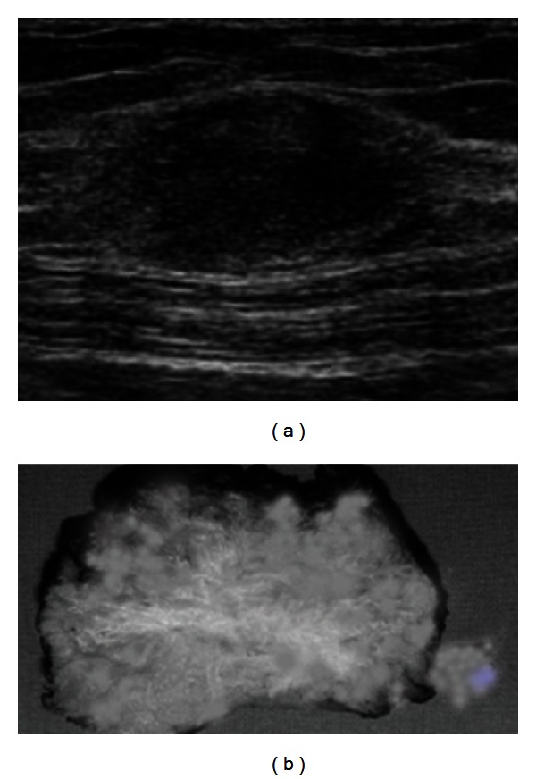
(a) USG in the transverse plane showing echogenic subcutaneous mass. (b) Gross photograph showing grey-white fibrous area with tiny cysts in the subcutaneous fat.

(a) USG in the transverse plane showing echogenic subcutaneous mass. (b) Gross photograph showing grey-white fibrous area with tiny cysts in the subcutaneous fat.