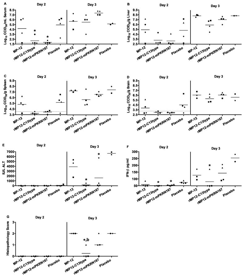
Figure 6. Viral titers, serum ALT, IFN-β, and histopathology in mice challenged s.c. with wt RVFV and vaccinated with MP-12 or MP-12 lacking NSs at 30 minutes post-exposure.
Animals were treated as described in Figure 5 and sacrificed on day 1 (not shown), 2 or 3 post-infection for analysis of A) serum, B) liver, C) spleen, and D) brain virus titers, E) serum ALT, F) serum IFN-β levels, and G) histopathology of the liver. The gray hashed lines indicate the limits of detection. Unique symbols on each day of sacrifice represent values for the same animal across all parameters. Due to death prior to time of sacrifice, data for several animals in the MP-12 and placebo groups could not be obtained. *P < 0.05 compared to animals receiving placebo, bP < 0.01 compared to animals receiving MP-12.

