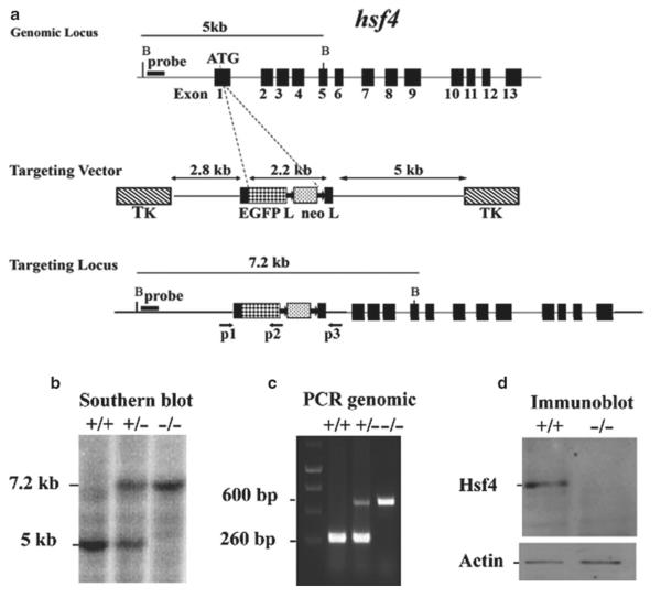
Fig. 5.
Targeted disruption of the hsf4 gene. (a) Wild-type hsf4 locus, targeting vector, and the predicted targeted allele following homologous recombination are shown. Exons are presented by black boxes. The probe used for Southern blotting and PCR primers P1, P2, and P3 are indicated by arrows below the targeted allele (16). (b) Bgl II-digested tail DNA (10 μg) from wild-type (+/+), hsf4+/− (+/−), or hsf4−/− (−/−) mice was hybridized with an external probe to yield bands of 5 and 7.2 kb for the wild-type and targeted hsf4 loci, correspondingly. (c) PCR-based genotyping assay amplifies fragments of 260 and 600 bp for wild-type and targeted hsf4 allele, respectively. (d) 50 μg protein from lens extracts of wild-type (+/+) or hsf4−/− (−/−) mice at P28 was analyzed by Western blotting using antibody specific to Hsf4b. As a control for equal protein loading, the blot was probed for β-actin.

