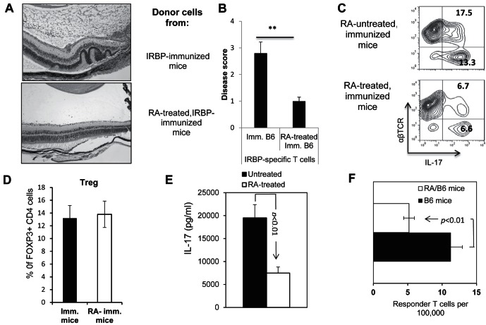
Figure 1.
ATRA-treated B6 mice generate decreased numbers of Th17 autoreactive T cells after immunization. (A, B) Splenic T cells from IRBP1-20/CFA-immunized B6 mice with or without ATRA treatment (200 mM, IP on day −3 and day 0) were enriched and stimulated for 48 hours with an optimal dose of immunizing peptide (10 μg/mL) under Th17 polarizing conditions. Then, the activated T cells were separated by Ficoll gradient centrifugation on day 3 and transferred adoptively to syngeneic naïve B6 mice. (A) The pathology of a representative eye section from each group. (B) Summarized the disease score results from three independent studies, each with 5 mice per group. (C) Cytoplasmic staining of in vitro activated IRBP-specific T cells. Using the protocol described for (A, B), on day 5 after in vitro stimulation with the immunizing peptide, the activated T cells were separated by Ficoll gradient centrifugation, and stained intracellularly with PE-conjugated anti-αβTCR antibodies and FITC-conjugated anti-IL-17 antibodies, followed by FACS analysis. (D) Foxp3+ among responder T cells of RA-treated and nontreated, immunized mice. (E) ELISA assay. The culture supernatant from immunized splenic T cells from ATRA-treated or untreated mice was tested by ELISA for IL-17 after 48 hours of stimulation with the immunizing peptide IRBP1-20. (F) Responder T-cell numbers were evaluated by LDA as detailed in the Materials and Methods. The results shown are representative of those from >5 experiments. ** P < 0.01, statistically significant.

