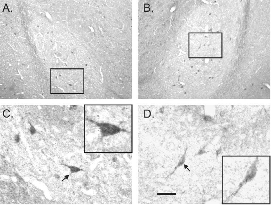
Figure 3.
Photomicrographs in A,B) and C,D) represent low and high magnification, respectively, images of immunohistochemical localization of the A-II type 1 receptor (AT1R) in the left and right basolateral amygdala (BLA: which is made up of the lateral amygdaloid nucleus (LA) and basolateral amygdaloid nucleus (BL)) at approximately –2.12 mm bregma) of an adult male rat. Black box inserts in A and B, represent high magnification photomicrographs shown in C and D, respectively. Black box inserts in C and D are higher magnification images of neurons in C and D that are indicated by arrows. Scale bars: 200 µm, A,B; 50 µm, C,D. Additional abbreviations: ec, external capsule; opt, optic tract.

