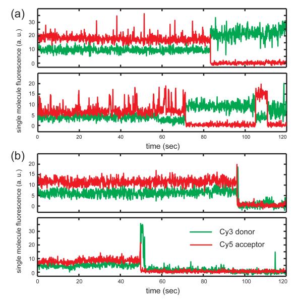
Figure 6. The smFRET pattern as a function of GTP concentration using a properly assembled (gp41)6-gp61-DNA primosome complex in a sequential two-step injection.
The experiments were performed using the same concentrations of gp41 (0.3 JM) and gp61 (50 nM), but varying GTP concentrations. Note that the donor fluorophore lies within the lagging (displaced) strand, and the acceptor within the leading strand. (a) Representative traces of a molecule exhibiting ‘incomplete’ unwinding FRET pattern. Following a high-to-low FRET conversion event, the donor signals for the lagging strand remained on the surface and also within the Förster distance of the acceptor, exhibiting a pattern reflecting repeated partial helicase unwinding events followed by rapid rewinding (reannealing) of the duplex portion of the fork construct. At a low concentration of GTP (6 JM) with a properly assembled (gp41)6-gp61-DNA primosome complex, ~83% of the fork constructs that we observed showed FRET conversion events with this incomplete unwinding pattern. (b) Representative traces of a fork construct exhibiting ‘complete’ unwinding FRET pattern, leading to a donor signal on the lagging strand that disappears abruptly right after the high-to-low FRET conversion event.

