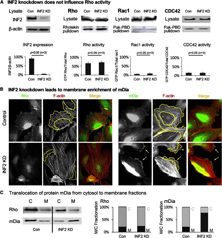
Figure 2.
INF2 counteracts mDia in maintaining cortical actin dynamics independent of Rho. (A) Rho/Rac/CDC42 activity in podocytes (undifferentiated) with or without INF2 KD was measured by pulldown assay. Expression of INF2 in cells was illustrated by immunoblotting and normalized to the protein level of β-actin. Cell lysates were incubated with Rhotekin or Pak-PBD–conjugated agarose, and the precipitates were subjected to immunoblotting using anti-Rho for Rhotekin pulldown or anti-Rac/CDC42 for Pak-PBD pulldown. The fraction of active (GTP bound, pulldown) Rho/Rac/CDC42 was compared as a percentage of the total Rho/rac/CDC42 level. (B) Podocytes with or without INF2 KD were costained with anti-Rho/anti-mDia and phalloidin. The changes in peripheral membrane recruitment of Rho/mDia (green) in association with actin filaments (red) were compared. Cortical actin area is circled by yellow lines. Local aggregation of mDia and retraction of lamellipodia in cells with INF2 depletion was highlighted by arrows. (C) Rho and mDia in the cytosolic and membrane fractions of cells were detected by Western blotting, and the membrane/cytosolic (M/C) fractions of Rho and mDia were compared in cells with or without INF2 knockdown. HSP 70 and Na+/K+ ATPase were used as markers for cytosolic and membrane fractions, respectively.

