Figure 1.

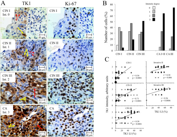
TK1 and Ki-67 immunohistochemistry staining of CIN and invasive cervical carcinomas. A) Example of TK1 and Ki-67 immunohistochemistry staining of CIN I, CIN II, CIN III and invasive cervical carcinomas (CA). The intensity (Int.) of the TK1 staining was scored as 0, 1, 2, and 3. Red arrows indicate cells with cytoplasmic TK1 staining only; yellow arrows indicate cells with both cytoplasmic and nuclear TK1 staining. Magnification x 400. B) Relative number of CIN and invasive cervical carcinoma patients with various TK1 staining intensities. C) Correlation between TK1 LI and TK1 intensity of CIN I to III lesions and of invasive cervical carcinomas with pathological stages II and III. Pearson-correlation statistical values are shown (r = regression coefficient).
