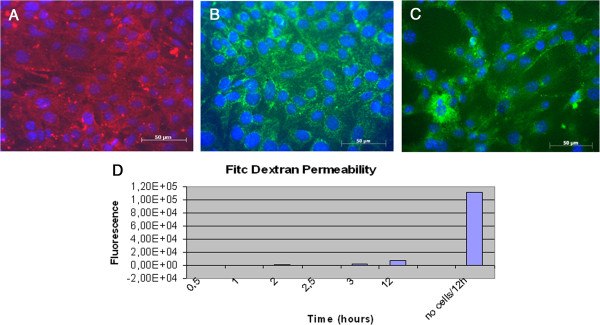
Figure 3.

Tight junctions markers and Vascular Permeability Assay of BMVECs. Murine BMVECs were isolated and cultured in EndoPM medium, then incubated 5 days in EndoPM medium without Hormone Mix. Immunofluorescent staining of confluent human brain microvascular endothelial cell monolayers for VE-cadherin (A), Claudin-5 (B), β-catenin (C). Nuclei were stained with DAPI. Scale bar = 50 μm. D) FITC-dextran permeability of the endothelium monolayer formed after isolation of BMVECs by means of the described protocol. Cells cultured in EndoPM medium and then plated in appropriate transwells strongly reduce macromolecules permeability throughout the barrier over 12 hours, as functional test of vascular permeability. All the experiments were repeated at passages n. 5, 10 and 15 without significant differences in the obtained results.
