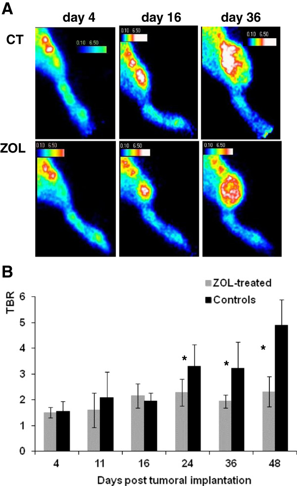
Figure 3.
99mTc-NTP 15-5 imaging of control and ZOL-treated groups. (A) Representative in vivo scintigraphic images of the tumor-bearing hindlimb obtained for controls and ZOL-treated animals at various stages of the study. (B) Semi-quantitative analysis of 99mTc-NTP 15-5 imaging: results are expressed as mean TBR values ± standard deviation. CT, controls; ZOL, zoledronic acid-treated; TBR, target-to-background ratio. The asterisk indicates p < 0.05.

