
Figure 5.
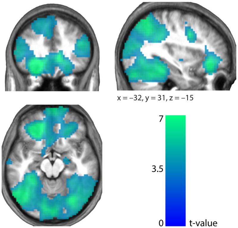
Stimulus category coding. In the frontal lobe, the central OFC (peak (x, y, z =−21, 38, −11), t = 11.14), the mFPC (peak (x, y, z = 6, 65, −11), t = 6.89) and the dorsolateral PFC (peak (x, y, z = −60, 17, 14), t = 11.34) contained distributed neural patterns pertaining to the identity of the stimulus under consideration. Toward the posterior, regions of the temporal lobes including the fusiform, inferior temporal and parahippocampal gyri, and areas around the intraparietal sulci also reflected category-discriminating activity (Supplementary Table 1). Results are presented at P < 0.005 FDR.