
Fig. 3. Immunocytochemical detection of phosphorylated GRK7 in HEK-293 cells.
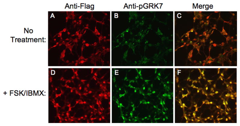
Cells were incubated in the absence (A, B, C) or presence (D, E, F) of 50 μM FSK and 1 mM IBMX for 30 min. Cells were stained simultaneously with anti-Flag (A, D) and anti-pGRK7 (B, E) antibodies at 1:1,500 and 1:100, respectively. Panel C represents the merging of images A and B. Panel F represents the merging of images D and E.