
Fig. 4. Detection of phosphorylated GRK7 in cones of pig retinas stained with anti-pGRK7.
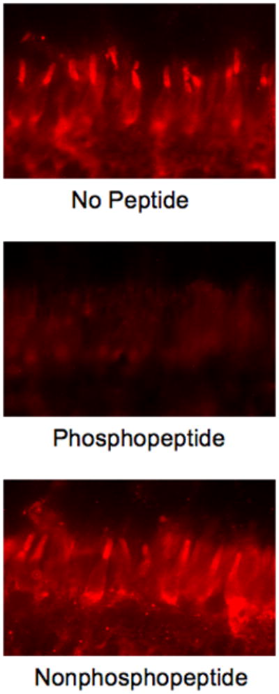
Anti-pGRK7 was preincubated at a 1:25 dilution overnight at 4 °C in the absence of peptide (upper panel), or with a 10-fold excess by weight of the phosphopeptide (middle panel), or the nonphosphopeptide (lower panel), followed by incubation with sections as described in the Materials and Methods. COS, cone outer segments, CIS, cone inner segments, ONL, outer nuclear layer.