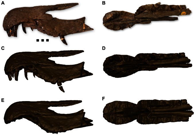
Figure 2. Lateral and ventral views of Baryonyx walkeri (NHMUK VP R9951) through the stages of digital preparation.
(A) The original specimen in left lateral view, (B) the original specimen in ventral view, (C) the digitally prepared original in left lateral view, (D) the digitally prepared original in ventral view, (E) final specimen with teeth removed and alveoli levelled, (F) final specimen with teeth removed and alveoli levelled showing cloned right maxilla. See Video S1 and S2 for more detailed visualisations of the preparation and reconstruction. Scale bar = 5 cm.

