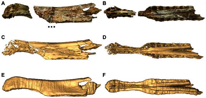
Figure 3. The digital preparation of Spinosaurus indet.
(NHMUK 16665) in lateral and ventral views. The original specimen – lateral view (A), and ventral view (B). The digitally prepared specimen with no matrix – lateral view (C), and ventral view (D). The rostral reconstruction is based on other specimens of Spinosaurus (e.g. [28]) and the B. walkeri rostra - lateral view (E) and ventral view (F). Video S3 and S4 for more detailed visualisations of preparation and reconstruction. Scale bar = 5 cm.

