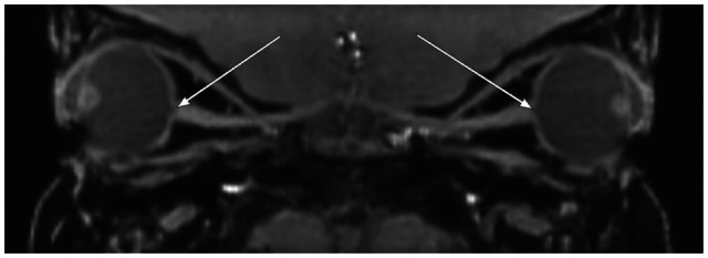
Figure 6.

Sagittal reformation of thin section T1-weighted orbital images demonstrating enlarged cup-to-disk ratios bilaterally with optic nerve excavation.
Notes: Arrows depict enlarged bilateral cup-to-disk ratios with optic nerve excavation. Buphthalmos, deep anterior chambers, and lens rounding are also depicted.
