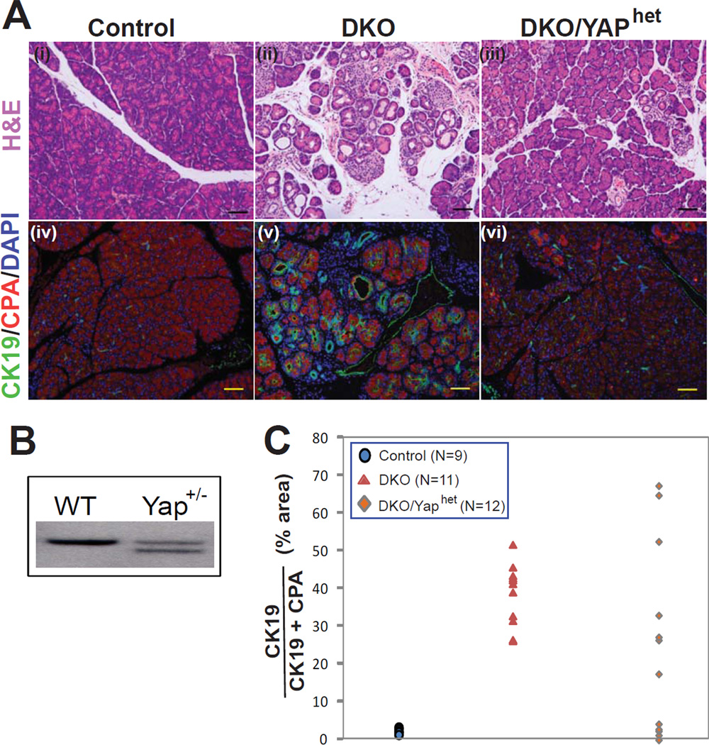
Figure 4. Yap lies downstream of Mst1/2 in the pancreas.
(A) H&E and immunofluorescence images of P30 control and DKO pancreata showing ductal metaplasia and enhanced CK19 staining (a b d e). In a subset of animals carrying one mutant Yap allele, pancreatic histology was normalized despite the absence of Mst1 and Mst2 (c f).
(B) Southern blot showing targeting of one Yap allele in embryonic stem cells.
(C) Quantification of ductal metaplasia. The area occupied by CK19+ cells was determined as a percentage of total area occupied by CK19+ and CPA1+ cells and plotted. In control animals, CK19+ cells occupied less than 5%, whereas in DKO animals CK19+ cells occupied more than 25%. Four DKO/Yaphet animals had a complete reversal of metaplasia. Scale bar: 100 µM.

