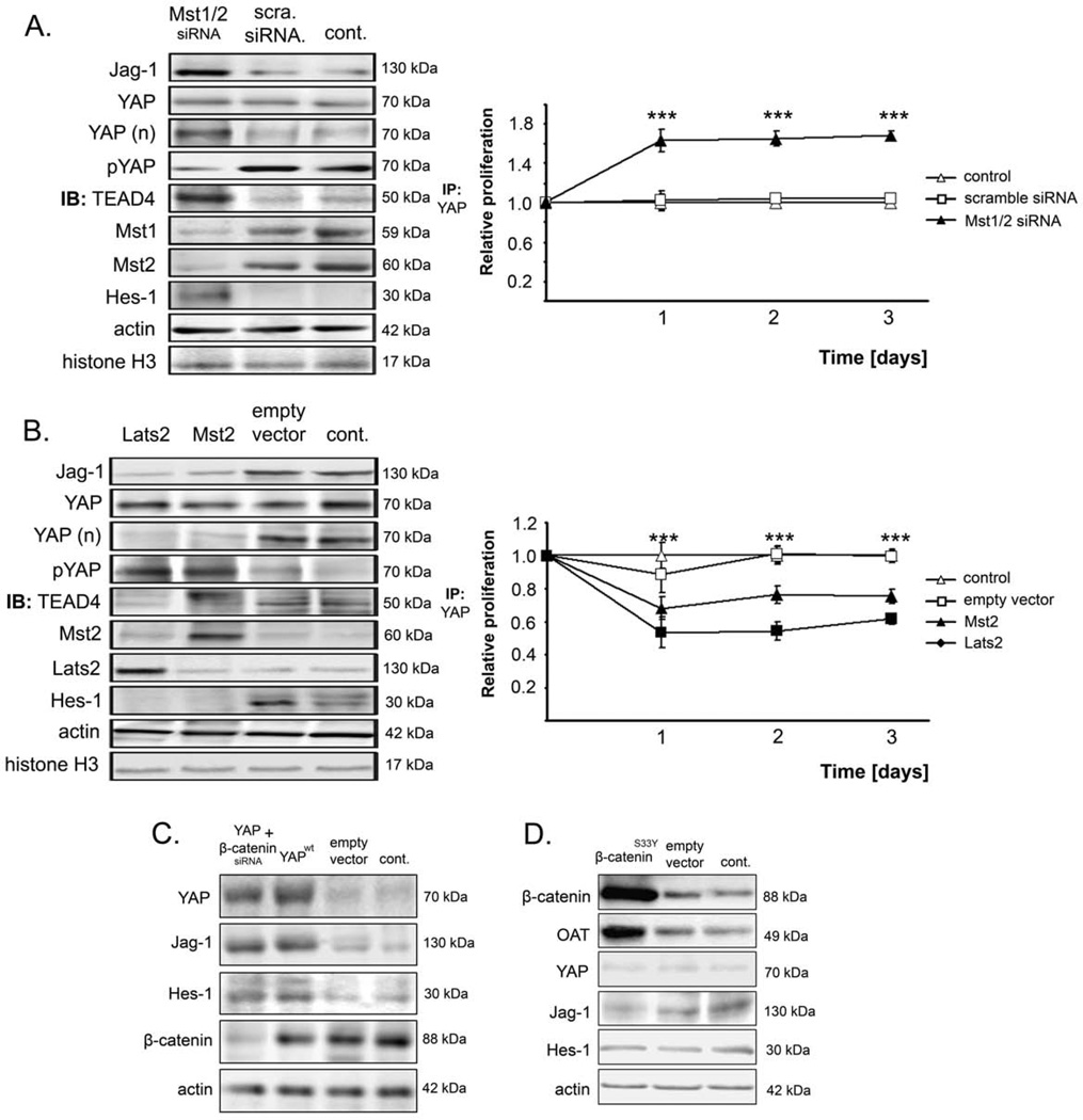
Figure 5. YAP regulates Jag-1 in a β-catenin-independent and MST/Latsdependent manner.
(A) Analyses of Jag-1, total YAP, nuclear YAP, phospho-YAP, TEAD4 (after IP with YAP antibody), Mst1, Mst2, and Hes-1 and corresponding relative proliferation of SNU-449 cells after combined inhibition of Mst1 and Mst2. SNU-449 cells were chosen because they show high-level expression of Mst1 and Mst2. Statistical analysis compares scramble siRNA and Mst1/2 siRNA. (B) Protein analyses of Jag-1, total YAP, nuclear YAP (after fractionation), phosphorylated YAP, TEAD4 (after IP with YAP antibody), Mst2, Lats2, and Hes-1 and corresponding cell proliferation of HuH-1 cells (showing low-levels of Mst2 and Lats2) after expression of Lats2 or Mst2. Statistical testing compares empty vector and Mst2 or Lats2, respectively. (C) Western blot analysis of YAP, Jag-1, Hes-1, and β-catenin in PLC/PRF/5 cells after overexpression of YAPwt and concomitant inhibition of β-catenin. (D) Analysis of β-catenin, OT, YAP, Jag-1, and Hes-1 in PLC/PRF/5 after overexpression of the constitutively active β-cateninS33Y mutant. For all Western blots, untreated (control), scramble siRNA-transfected, or empty vector-transfected cells were used as controls. Actin served as loading control. Values represent means ± SD. For statistical comparison, the Mann-Whitney U test was used. ***p<0.001.

