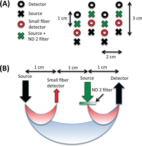
Figure 1.
NIRS probe. (A) Geometry (B) Sensitivity In order to avoid detector saturation, 200 μm-core fibers were used for the SS detectors (shown in red) and a piece of optical filter (Kodak WRATTEN ND 2.00) was glued at the tips of standard NIRS fibers (shown in green) for the additional sources of the SS measurements.

