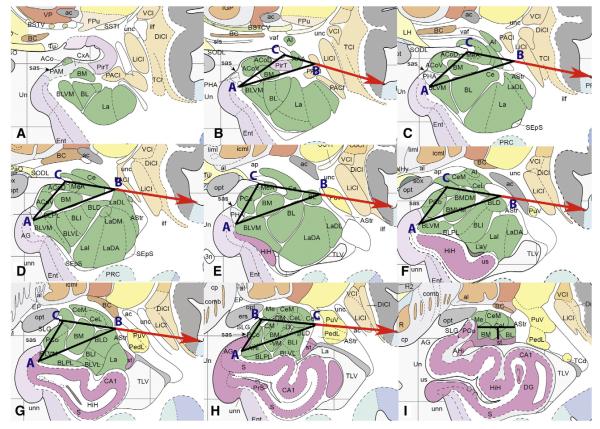
Fig. 3.
Detail of geometrically determined protocol for segmenting the amygdala, from most rostral (A) to most caudal (I). See text for explanation of points A, B, and C. A is not segmented because the whole amygdala tracing is labeled as BL at this level. Horizontal line from Point C to Point B extends beyond boundaries of whole amygdala to indicate direction toward the circular sulcus, but ends at Point B in the protocol. I is split evenly on the horizontal axis and on the vertical axis below the level of the central nucleus. See Box 1 for abbreviations.
Images are reproduced with permission from Elsevier Inc.

