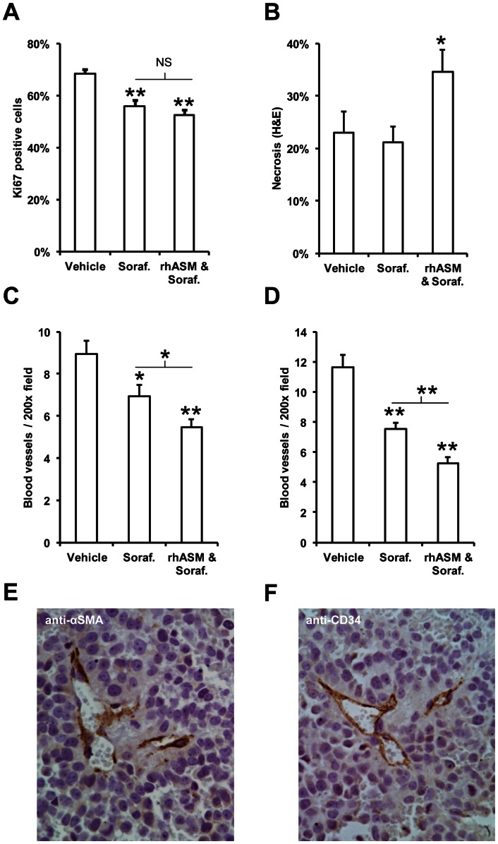
Figure 3. rhASM/sorafenib co-treatment reduces blood vessel density and increases necrosis in Huh7 tumors.
(A) Mean number of Ki67 positive cells in tumors from mice treated with sorafenib (Dunnett's post-hoc p<0.005) and with rhASM/sorafenib combination (Dunnett's post-hoc p<0.001) was significantly lower than vehicle (ANOVA df (2,30), F = 14.63, p<0.001). No significant difference was observed between Ki67 staining in tumors from sorafenib and rhASM/sorafenib treated mice (t = 1.19, df 22, p = 0.249). (B) Necrosis was significantly increased (ANOVA, df (2,30), F = 3.66, p = 0.038) in rhASM/sorafenib treated mice (Dunnett's post hoc > control, p = 0.043) compared to vehicle treated mice, while sorafenib treated mice were not different from control (Dunnett's post hoc > control, p = 0.760). Necrosis in rhASM/sorafenib treated mice was significantly greater than in mice treated with sorafenib alone (t = −2.39, df (22), p = 0.26). (C) Anti-αSMA blood vessel staining revealed a significantly lower number of vessels (ANOVA df (2,30), F = 12.57, p<0.005) in sorafenib (Dunnett's post hoc test p = 0.020) and rhASM/sorafenib treated mice (Dunnett's post hoc test p<0.001). The rhASM/sorafenib group also had significantly less αSMA blood vessels than the sorafenib group (t = 2.25, df (22), p = 0.031). (D) Anti-CD34 blood vessel staining confirmed the αSMA results, showing a significantly lower number of vessels (ANOVA df (2,30), F = 32.07, p<0.001) in sorafenib (Dunnett's post hoc test p<0.001) and rhASM/sorafenib treated mice (Dunnett's post hoc test p<0.001). The combined rhASM/sorafenib group had significantly less CD-34 positive blood vessels than the sorafenib group (t = 3.56, df (22), p = 0.002). Selective staining of blood vessels in paraffin embedded tumor sections stained is shown with anti- αSMA (E) and anti-CD34 markers (F). *p<0.05, **p<0.001.

