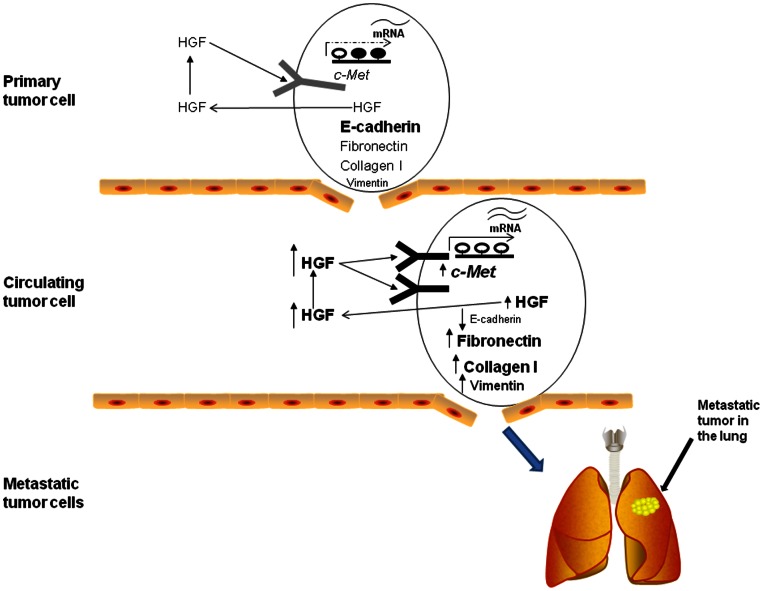
Figure 7. Summary of potential role of HGF/c-Met in hematogenous dissemination of HCC.
Schematic diagram illustrating the role of HGF/c-Met in hematogenous dissemination of HCC cells. Primary tumor cells express HGF and c-Met with partial promoter demethylation. This is associated with good expression of E-cadherin and limited expression of fibronectin, collagen I and vimentin. However, when HCC cells escape from the liver and start circulating in the blood-stream, increased expression of HGF and c-Met associated with increased promoter demethylation is observed. Circulating HCC cells also display evidence of EMT: loss of E-cadherin, increased fibronectin, increased collagen I and increased vimentin expression. We conclude that increased HGF and c-Met may induce EMT in CTCs to sustain hematogenous dissemination.

