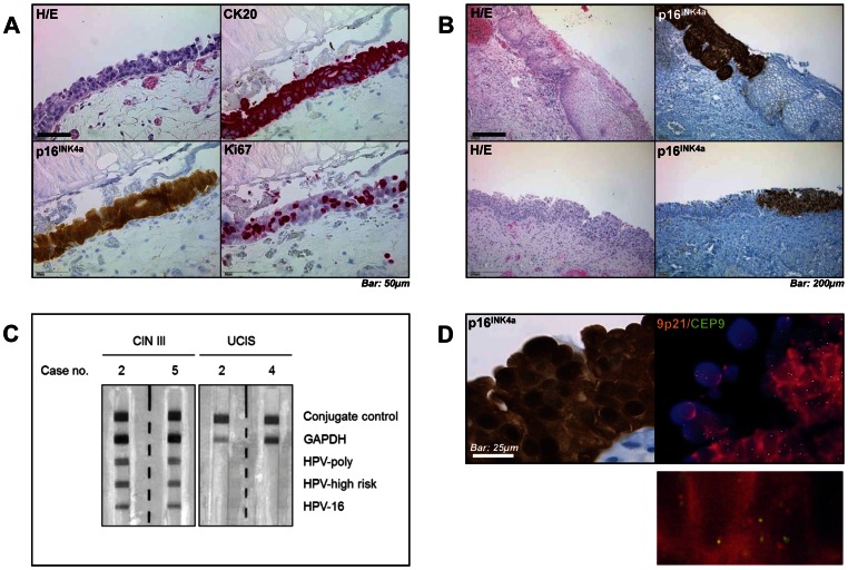
Figure 1. Immunohistochemical characterization, HPV testing and FISH analysis in UCIS.
A, Histologic microphotograph of urothelial carcinoma in situ (UCIS) shows loss of nuclear stratification, positivity for Cytokeratin 20 and p16INK4a as well as increased proliferative activity as indicated by Ki-67 immunostaining. B, Comparison between high-grade cervical intraepithelial neoplasia (CIN III, upper row) and UCIS (lower row) shows a comparable distribution of p16INK4a immunopositivity. C, Reverse hybridization blotting detects high-risk human papillomavirus (HPV) genotype 16 DNA in representative CIN III, but not in UCIS samples. D, Fluorescence-in-situ-hybridization (FISH) analysis shows no amplification of CDKN2A gene encoding for p16INK4a (orange) compared to control (centromere 9, green) in representative p16INK4a-positive UCIS. Scale bars as indicated.

