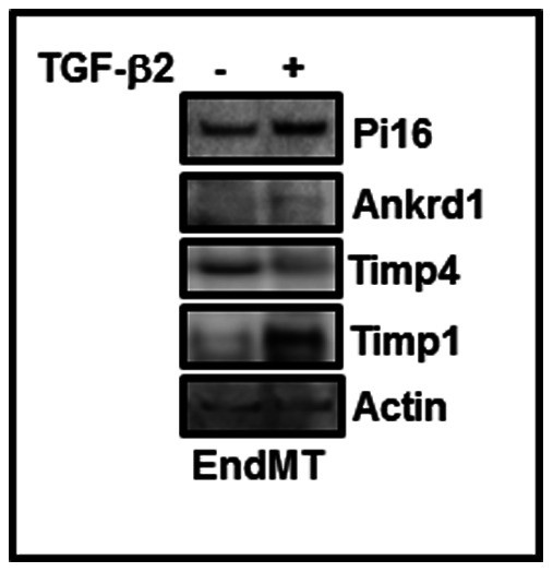
Figure 6. Levels of Ankrd1, Pi16, Timp4 and Timp1 in TGF-β-treated endothelial cells.

Mouse cardiac endothelial cells were treated with TGF-ß2 (10 ng/ml) for 7 days. At the end of incubation, control and EndMT-derived fibroblast-like cells were harvested and whole lysates were prepared. Equal amounts of proteins were subjected to Western blot using specific antibodies as indicated.
