
Fig. 3.
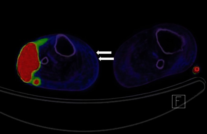
The positron emission tomography-computed tomography of the fludeoxyglucose-avid malignant tumor in the upper lateral aspect of the right lower leg with possible extension to the adjacent soft tissue and a unilateral subcutaneous honeycomb-appearance (white arrows) suggests long-standing lymphedema. There was no evidence of metastasis.