Fig. 4.

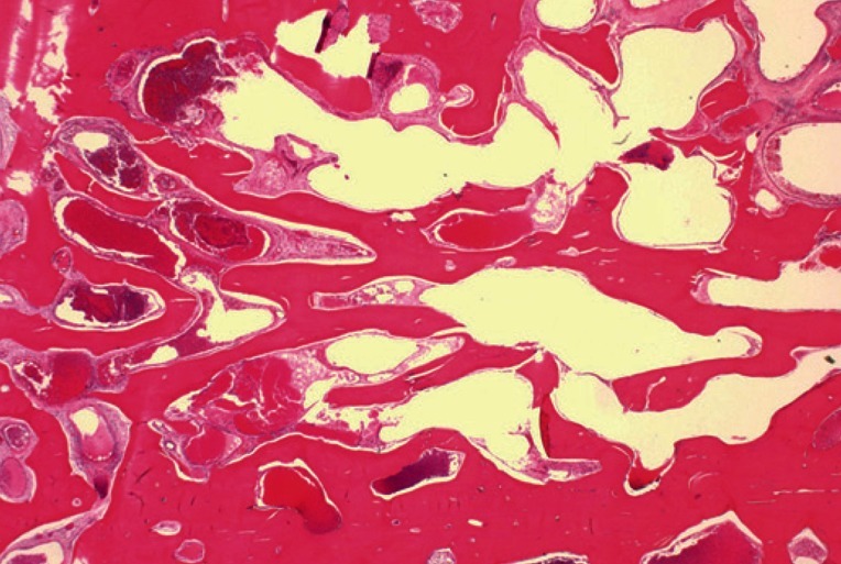
Microscopic findings with H&E stain: cavernous hemangioma showing extended, thin-walled vessels, and sinuses lined by a single layer of endothelial cells (×12).

Microscopic findings with H&E stain: cavernous hemangioma showing extended, thin-walled vessels, and sinuses lined by a single layer of endothelial cells (×12).