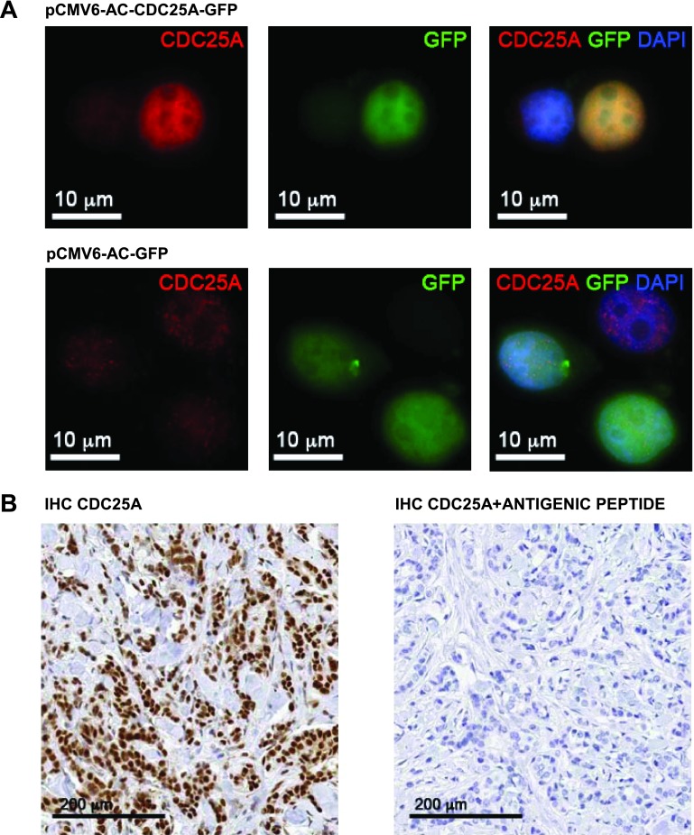
Figure 1.
(A) CDC25A (144) sc-97 antibody (Santa Cruz Biotechnology) validation on the breast cancer cell line MCF-7. Immunofluorescence with CDC25A antibody in the MCF-7 breast cancer cell line transfected with either pCMV-AC-CDC25A-GFP or pCMV-AC-GFP: The antibody detected a strong CDC25A expression in the CDC25A-GFP-transfected MCF-7 cells compared to the pCMV-AC-GFP-transfected MCF-7 cells. DAPI counterstaining underlined the nuclear localization of CDC25A (red signal, CDC25A; green signal, GFP; blue signal, DAPI; yellow signal, merge). (B) CDC25A (144) sc-97 antibody IHC validation on FFPE breast cancer tissue samples. CDC25A nuclear localization was detected by IHC on FFPE breast cancer tissue sample (left); CDC25A antibody preabsorption testing using the antigenic peptide (CDC25A 144 P sc-97P; Santa Cruz Biotechnology) showed a complete block in the immunoreactivity (right).

