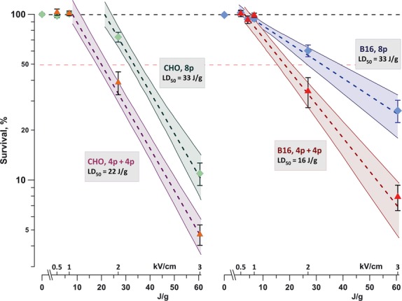
Fig. 1.

Enhancement of the cytotoxic effect of 100-μs electric pulses (EP) by exposure fractionation. CHO cells (left panel) and B16 cells (right panel) were exposed to eight pulses (100 Hz) delivered either as a single dose (8p) or a split dose (4p+4p) with 5-min. interval between two trains. The graphs show cell survival (mean ± SE for three to six independent experiments) versus the dose for different EP treatments. Dashed lines are the best fit data approximations using exponential function; shaded areas denote 95% confidence intervals. Cell survival was measured by MTT assay at 24 hrs post exposure. Legends show lethal dose values for elimination of 50% of cells (LD50) by the respective exposure protocols.
