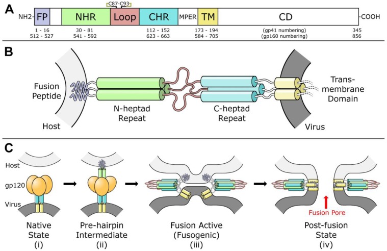
Figure 1.
(A) Diagram of fusion protein gp41 sequence. From the N-terminus, the fusion peptide (FP), N-heptad repeat (NHR), loop region, C-heptad repeat (CHR), membrane-proximal external region (MPER), transmembrane domain (TM), and cytoplasmic domain (CD) are labeled. A disulfide bond between Cys 87 and Cys 93 in the loop region is indicated. (B) Model of gp41 trimer in the pre-hairpin intermediate conformation. In this model, gp41 spans from the host membrane (light gray) to the viral membrane (dark grey). Regions are colored according to the diagram in part (A). Cytoplasmic domain is omitted. (C) Model for gp41-mediated membrane fusion. In the native state (i) and the pre-hairpin intermediate (ii), gp120 receptors and co-receptors are omitted for clarity. In the fusion active state (iii) and the post-fusion state (iv), gp120 is omitted for clarity, and a second six-helix bundle is shown to illustrate cooperativity in forming the fusion pore. Red arrow indicates fusion pore. Concept for Figure adapted from Chan et al. [27].

