
Figure 3.
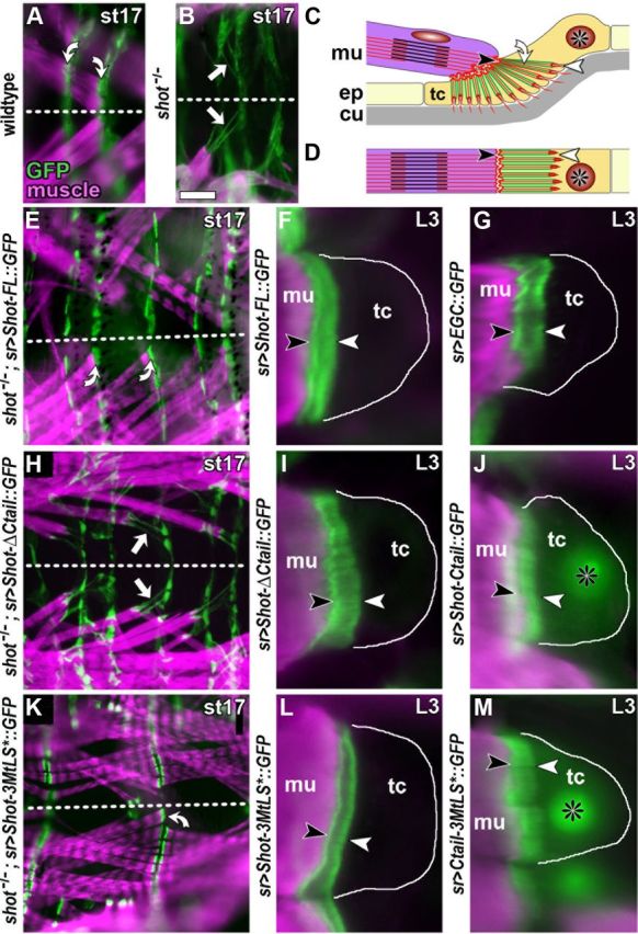
Localization and rescue capability of mutant Shot constructs in tendon cells. A, A late stage 17 (st17) wild-type embryo in plain view (anterior left; dashed line indicates midline) displaying muscles (stained with phalloidin, magenta) which attach with their tips to tendon cells with prominent cytoskeletal arrays (curved arrows; stained with actin::GFP, green). B, In shot−/− mutant embryos, tendon cell integrity is affected as reflected by abnormal elongation of actin::GFP-labeled cytoskeletal arrays (white arrows). C, D, Diagrams illustrating tendon cell morphology in lateral (C) and plain (D) view; muscles (mu, magenta) attach to basal surfaces (black arrowheads) of tendon cells (tc; asterisks indicate nuclei), which are specialized cells of the epidermis (ep); apical tendon cell surfaces (white arrowhead) link to the exoskeleton called cuticle (cu, gray); apical and basal tendon cell surfaces are connected through cytoskeletal arrays (curved arrow) which are composed of parallel actin fibers (red) and MTs (green) and appear as a continuous band in horizontal view (D). E, H, K, Plain views of shot−/− mutant embryos with targeted expression of Shot-FL, Shot-ΔCtail or Shot-3MtLS (as indicated on the left); successful rescue of tendon cell integrity by Shot-FL and Shot-3MtLS is indicated by curved arrows in E and K, respectively; arrows point at stretched cytoskeletal arrays reflecting failed rescue through Shot-ΔCtail; a similar lack of rescue was observed for Shot-ΔGRD (Bottenberg et al., 2009). F, G, I, J, L, M, Images from late L3 larvae show muscle tips (magenta) attached to tendon cells (outlined with white line) which express different GFP-tagged constructs (green; as indicated on the left of each panel; all symbols and abbreviations as in C); Shot-FL, Shot-ΔCtail, Shot-3MtLS* and EGC all show strong association with cytoskeletal arrays with a slightly higher concentration at apical and basal ends. Strong MT association is surprising especially for Shot-ΔCtail, but similar observations were made for Shot-ΔGRD (Bottenberg et al., 2009); they might be explained through dimerization of these deletion constructs with endogenous Shot or interactions of N-terminal or central domains with other constituents proteins of cytoskeletal arrays. In contrast, Ctail and Ctail-3MtLS* show weaker and homogeneous localization at cytoskeletal arrays, and higher cytoplasmic and nuclear levels. Scale bar (in A) A, B, E, H, K, 40 μm; F, G, I, J, L, M, 7 μm.