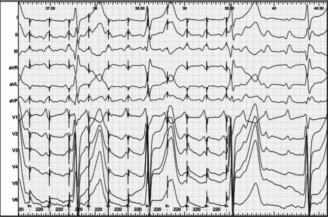
Figure 10.

Entrainment with fusion as demonstrated by stimulation after P-wave onset. This 12-lead ECG tracing shows the last 11 beats of a pacing train at a cycle length of 220 ms delivered during an atypical flutter with a cycle length of 240 ms. The tracing is considerably gained to show P-wave morphology. Please note that each stimulus artifact is delivered after the P-wave onset, during its inscription. Capture of each stimulus can be deducted by some deformation in the P-wave contour as compared with the flutter P wave, but most importantly because the P-wave cycle length is accelerated to the pacing cycle length. Thus, since each stimulus captures but is delivered after the P-wave onset, there must be fusion, and it can be assured that fusion is due to an exiting wavefront from the tachycardia itself.
