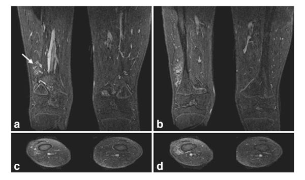
Figure 2.
Single 1.1-mm-thick coronal and axial slices from the high spatial resolution, nonsubtracted late phase image from Patient 2. Single coronal slices (a,b, anterior to posterior) and axial slices (c,d, superior to inferior) show the fine detail of the vascular malformation located in the right vastus intermedius with no bony involvement. Enlarged draining veins (arrow) are shown in (a). The 3D volume imaged in this late phase scan is the same as that for the CAPR time series.

